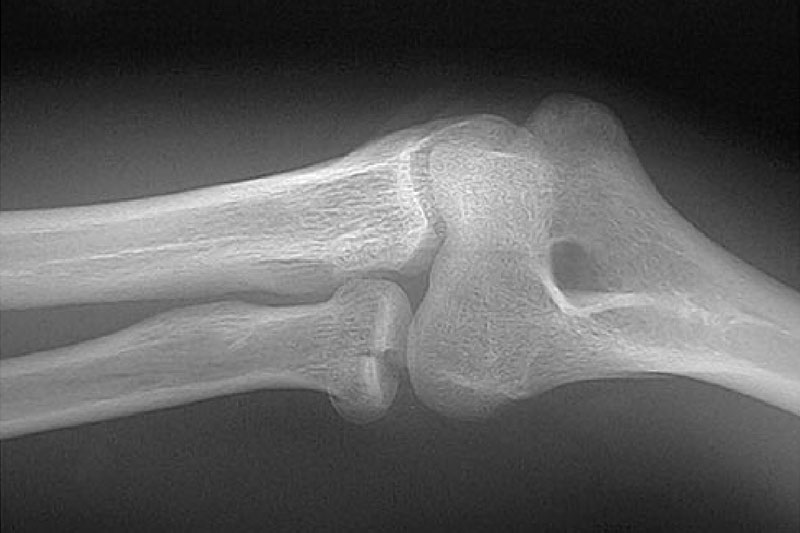
Frattura del capitello radiale
Among all the fractures of the elbow, the radial head appears involved in 20% of the cases. This type of fracture is generally caused by indirect traumas, because of the fall on the upper limb with hyperextended hand and extended elbow.
Acute pain can lead to a limitation of motion and strength.
In addition to a standard radiography, in some cases oblique projections can be useful for the best view of the radial head. MRI is the most sensitive test for diagnosis.
The compound fractures are treated with a rigid brace for about 10 days. The beginning of the rehabilitation treatment, with a cautious but premature mobilization, is essential in order to avoid the risk, which very often occurs, of rigidity.
Displaced fractures are often non-operatively treated with a splint for two weeks followed by mobilization only in flexion-extension. Then, after an x-ray inspection, the process of rehabilitation is taken to an end. The results are positive in 70% of the case.
Radial head fracture – Surgery
This is typically treated with the open-air reduction of the fracture and internal fixation.
The surgical trauma, added to the one produced by the injury would lead to a disabling joint stiffness, so it is essential to start early an appropriated rehabilitative treatment.
Radial head fracture – Rehabilitation
Among the elbow fractures, the radial head is interested in 20% of cases. The fracture is usually caused by indirect trauma due to the fall on the upper limb with hyperextended hand, extended elbow and forearm pronated.
Rehabilitation, immobilisation, surgery and the recovery time vary according to the location and the type of fracture (3 types), but all have a similar treatment protocol.
In the first phase of the rehabilitation program it is crucial a cautious but early mobilization of the elbow, to prevent the establishment of joint stiffness, early passive traction then progressively through active stretching and auto mobilization. In this first stage physical therapies (laser, iontophoresis) and ice can be used to relieve pain.
Once you have achieved an excellent joint mobility you can proceed to the second phase of the treatment protocol focused on the recovery of the strength of the muscle chain with strengthening exercises prone/supinators of the wrist, flexion/extensor of the forearm, biceps and triceps, shoulder stabilizers with weights and elastic in both concentric and in the eccentric.
The last phase of rehabilitation takes place in the field and its objective is the resumption of manual coordination through exercises launch/socket objects (neuromotor training) and the recovery of technical gestures with exercises to train the fall damping mode, for the prevention of re-injury.

